Showing posts with label 3D Image-Development. Show all posts

Friday, 9 December 2016

New Anatomy / Physiology, App

An Irish developed app has been listed by Apple as one of the 16 most innovative apps and games for 2016

Complete Anatomy, designed by Dublin-based 3D4Medical, beat competition from two million other apps that were published in Apple's App Store this year.
The app gives people learning anatomy or physiology a 3D view of the body, layer by layer, and allows the user to zoom, rotate, cut through structures and see behind layers.
Recordings can also be made for educational purposes, and content can be shared via the cloud.
The App Store Best of 2016 shortlist was picked by Apple's Editorial Team.
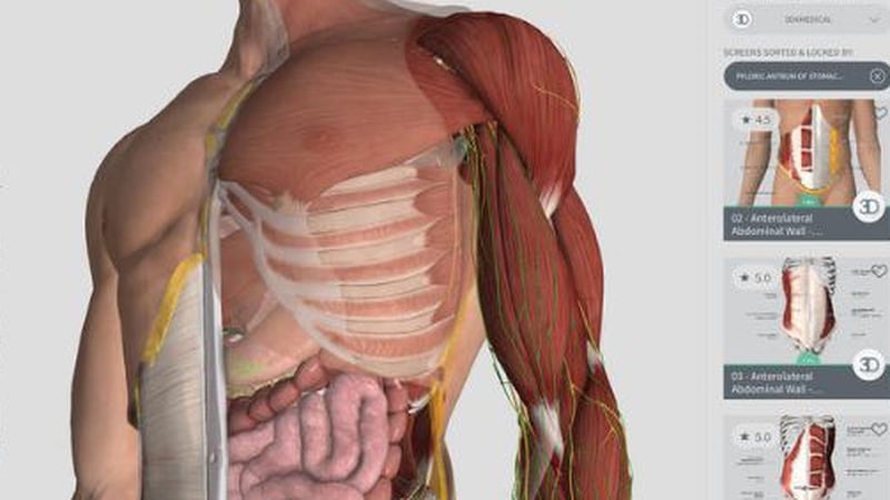
A screenshot of the 3D4Medical app from Apple iTunes Store
A screenshot of the 3D4Medical app from Apple

Last year Complete Anatomy received the Apple Design Award for its technical excellence and outstanding design and innovation, and was also featured on stage by Apple during a high-profile iPad product launch.
The app has reached the top of the app store medical charts in 148 countries, and the overall charts in the online shop in 52 states.
At the start of this year, 3D4Medical announced it was taking on 70 new staff as it bolstered its product development line and grew sales overseas.

Monday, 12 September 2016

3D Images - New Technique at TCD

A new bone-scanning technique that generates ultra-high resolution 3D images without the need for potentially harmful x-rays has been developed by scientists in Ireland.

The technique, devised by chemists at Trinity College Dublin and the Royal College of Surgeons in Ireland, involves the use of tiny pieces of gold, which have compounds attached to them that are luminescent in certain conditions.
The fragments are attracted to tiny fissures that appear when bones crack, and attach themselves to the area.
Using Magnetic Resonance Imaging (MRI), the researchers can then scan the damaged area which is revealed in a three-dimensional map of the microcracks.
The research, published in the journal Chem, could lead to the development of a method for diagnosing problems with bone strength and other degenerative bone diseases like osteoporosis.


The research could lead to the development of a method for diagnosing problems with bone strength

The technique also avoids the use of x-ray imaging, which has been associated with a higher risk of cancer.
"The nano-agent we have developed allows us to visualise the nature and the extent of the damage in a manner that wasn't previously possible," said TCD Professor of Chemistry, Thorri Gunnlaugsson.
"This is a major step forward in our endeavour to develop targeted contrast agents for bone diagnostics for use in clinical applications."
"Current x-ray techniques can tell us about the quantity of bone present but they do not give much information about bone quality," said RCSI Professor of Anatomy Clive Lee, who was also involved.
"By using our new nano-agent to label micro-cracks and detecting them with magnetic resonance imaging, we hope to measure both bone quantity and quality and identify those at greatest risk of fracture and institute appropriate therapy.
"Diagnosing weak bones before they break should therefore reduce the need for operations and implants - prevention is better than cure."