Showing posts with label RCSI. Show all posts

Thursday, 20 October 2016

New Technology to Address Bedsores!

New technology designed to eliminate the problem of painful, potentially dangerous and costly bedsores is being tested within Irish hospitals. 

The new wireless device is able to detect the pressure sores up to ten days, before they appear on the skin's surface, giving clinicians time to treat them before they become problematic.
Pressure ulcers are injuries to the skin and underlying tissue resulting from prolonged pressure on the skin, and most commonly develop on skin in bony areas of the body, like heels, ankles, hips and tailbone.
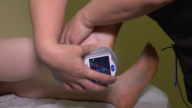

The handheld SEM Scanner, from California based Bruin Biometrics, detects increases in sub-epidermal moisture which is a biophysical marker of damaged skin.

Long-stay hospital patients who are confined to bed and people who suffer from reduced mobility, are at increased risk of developing the problem.
If left untreated they can become very painful, can lead to blood infection, and even death.
It is estimated that as well as causing great discomfort and distress to patients and their families, bedsores cost the Irish health service €250 million a year to manage.
Last year nearly one in ten serious medical events in Irish healthcare institutions was related to late-stage bedsores.
Traditionally pressure ulcers have been identified through visual inspection by care-givers, and treated using established protocols.
However, often by the time the sore is visually apparent and spotted, much damage has already been done, making it more difficult and time consuming to treat.
"The challenge with the visual assessment is that we can't see what is going on under the skin," said Professor Zena Moore, Head of School of Nursing and Midwifery at the Royal College of Surgeons in Ireland.

Monday, 12 September 2016

3D Images - New Technique at TCD

A new bone-scanning technique that generates ultra-high resolution 3D images without the need for potentially harmful x-rays has been developed by scientists in Ireland.

The technique, devised by chemists at Trinity College Dublin and the Royal College of Surgeons in Ireland, involves the use of tiny pieces of gold, which have compounds attached to them that are luminescent in certain conditions.
The fragments are attracted to tiny fissures that appear when bones crack, and attach themselves to the area.
Using Magnetic Resonance Imaging (MRI), the researchers can then scan the damaged area which is revealed in a three-dimensional map of the microcracks.
The research, published in the journal Chem, could lead to the development of a method for diagnosing problems with bone strength and other degenerative bone diseases like osteoporosis.


The research could lead to the development of a method for diagnosing problems with bone strength

The technique also avoids the use of x-ray imaging, which has been associated with a higher risk of cancer.
"The nano-agent we have developed allows us to visualise the nature and the extent of the damage in a manner that wasn't previously possible," said TCD Professor of Chemistry, Thorri Gunnlaugsson.
"This is a major step forward in our endeavour to develop targeted contrast agents for bone diagnostics for use in clinical applications."
"Current x-ray techniques can tell us about the quantity of bone present but they do not give much information about bone quality," said RCSI Professor of Anatomy Clive Lee, who was also involved.
"By using our new nano-agent to label micro-cracks and detecting them with magnetic resonance imaging, we hope to measure both bone quantity and quality and identify those at greatest risk of fracture and institute appropriate therapy.
"Diagnosing weak bones before they break should therefore reduce the need for operations and implants - prevention is better than cure."

Thursday, 19 March 2015

RCSI study: Do flip-flops increase the risk of tripping?

Flip-flops really do increase the risk of tripping, the results of a fresh study have confirmed.

Three researchers from the RCSI’s School of Medicine, the Gait Laboratory, Central Remedial Clinic (CRC), in Clontarf, and the School of Physiotherapy set out to answer the question ‘Do flip-flops increase the risk of tripping when walking?’
“From our research, we concluded that the required increase in knee flexion and ankle dorsiflexion, along with the sizeable difference between barefoot and flip-flops in ground clearance, may increase the risk of tripping during walking in flip-flops, compared to bare feet,” they determined.
Fifteen healthy participants between the ages of 17 and 75 years with no history of gait pathology were recruited for the research that took place in the Gait Laboratory of the CRC.
Light-emitting diode markers were placed on the hallux, base of the fifth metatarsal and heel of each foot, as well as the ankle and knee joints.
Plastic wands were then attached to the shanks, thighs and hips, to define the planes of movement along the lower limbs, with additional markers attached to corresponding locations on the slippers.
Each participant made five walks barefoot and five in flip-flops, for data collection.
The researchers said 12 key kinematic and temporal-spatial gait parameters were analysed using three-dimensional motion analysis: gait speed, single and double support duration, step length, total ankle range of motion (ROM) in the sagittal plane, total sub-talar joint ROM in the coronal plane, and peak knee flexion in swing.
Minimum toe clearance when barefoot and minimum shoe clearance in slippers in swing, knee and ankle position at initial contact and separation distance between barefoot and flip-flop markers, were also used.
They found that wearing slippers caused an increase in peak knee flexion in swing and ankle dorsiflexion.
The average separation between the heel and base of the flip-flop, in pre-swing, was 8.8cm (SD = 1.48). In mid-swing, the slipper cleared the ground by an average 1.6cm (SD = 0.56) at its lowest point, in contrast to the mean barefoot clearance of 4.2cm (SD = 0.8).

Monday, 16 February 2015

New research to guide GP management for patients with multiple chronic illnesses

The new research recommends providing continuity of care with one named GP where possible, prioritising the aspects of care that matter most to patients (such as difficulties in physical functioning), and providing regular reviews of prescriptions. The study, recently published in the British Journal of Medicine, was led by researchers at the HRB Centre for Primary Care Research in the Royal College of Surgeons in Ireland.

The paper entitled, ‘Managing patients with multimorbidity in primary care' can be viewed online on the BMJ website at http://www.bmj.com/content/350/bmj.h176

Current clinical guidelines, which recommend best practice for GPs, tend to focus on single conditions which make decision making more challenging for those patients with several co-existing conditions.

Commenting on this publication, Graham Love, Chief Executive at the Health Research Board said,
‘It is very important that care for people with multimorbidity is organised and delivered based on best research evidence in order to improve the outcomes that really matter to patients. This review offers guidance for GPs to help support this process.’

More information is available from the press release on the RCSI website.

Wednesday, 26 November 2014

Educational gaming App launched for surgeons

RCSI has launched a new gaming App for iPhone and iPad aimed at testing students’ and doctors’ medical knowledge on the go.
The SurgiQuiz game, which allows surgeons to obtain Continuous Professional Development (CPD) credits, is targeted at surgeons, health sciences students and anyone that is interested in human anatomy.
Developed by the College’s Department of Surgical Affairs in conjunction with the Department of Anatomy, it consists of 3,000 tough questions that are aimed at challenging even practising surgeons.
The game also includes a global leaderboard so people can compete to get their name on the board.
The free App is available for download from the App store on iPhone and iPad. An Android version is also in development.

Wednesday, 2 April 2014

Research may lead to a new bowel cancer detection method

HRB-funded research at the Royal College of Surgeons in Ireland, and just published in the European Journal of Clinical Microbiology and Infectious Diseases, could lead to a new biomarker for bowel cancer
Long, pointy Gram-negative rods typical of Fusobacterium nucleatum. Image from www.infectionnet.org

The research, led by Dr David Hughes at the Department of Physiology and Medical Physics, RCSI found a significantly increased presence of a common microbe, Fusobacterium nucleatum (Fn), in tissue and stool samples of patients with colorectal cancers and colorectal benign tumours.
Commenting on the findings, Dr Hughes said,
'Our research found that cancer patients with low bacterial levels had significantly longer survival times than patients with moderate and high levels of the bacterium. Also, for patients with a benign tumour, we found that the presence of the bacteria may be a risk factor for disease progression from tumour to cancer. This is a significant finding because it highlights the potential of Fn detection as a possible indicator of colorectal cancers'.

View full article here
Efforts to combat Fn infection could be considered for colorectal cancers patients with high levels of the bacterium to improve the survival prospects for these patients.
More details are available from the RCSI